

Pathology models are three - dimensional representations of human organs, tissues, or pathological conditions, crafted to replicate the anatomical structure and characteristics of real - world specimens. These models serve as invaluable educational and diagnostic aids, offering detailed visualizations that can enhance understanding and facilitate more informed decision - making. As a dedicated pathology model supplier, I've witnessed firsthand the growing interest in these tools and the debates surrounding their widespread use in clinical practice.
The Role of Pathology Models in Clinical Education
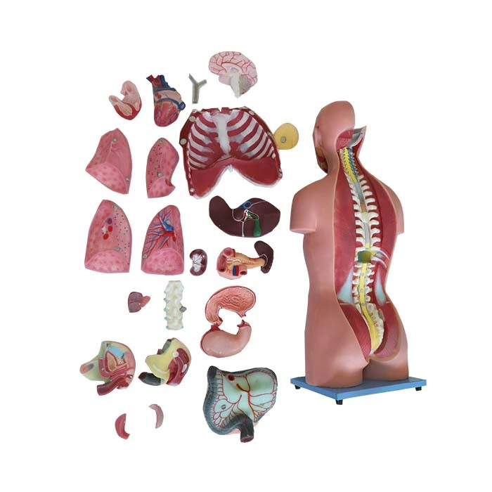
In the realm of medical education, pathology models have long been a staple. Medical students often rely on these models to supplement their learning of human anatomy and pathophysiology. For instance, a Model Of Human Body Showing Internal Organs can provide a tangible way for students to explore the complex relationships between different organs within the body. It allows them to visualize how various systems interact, which is crucial for understanding normal physiological functions and how they can be disrupted in disease states.
Pathology models also play a vital role in teaching surgical techniques. Surgeons - in - training can practice on these models to develop the necessary dexterity and spatial awareness required for complex procedures. For example, a Stomach Model Anatomy can be used to simulate gastric surgeries, enabling trainees to familiarize themselves with the anatomical landmarks and the steps involved in the operation. This hands - on experience is irreplaceable and can significantly improve the confidence and competence of future medical professionals.
Applications in Clinical Diagnosis
In clinical diagnosis, pathology models can serve as powerful visual aids. When communicating a patient's condition to other healthcare providers or to the patient themselves, a well - crafted model can convey information more effectively than a two - dimensional image or a verbal description. For example, in cases of congenital heart defects, a model can show the exact nature of the defect, such as the location and size of a ventricular septal defect. This can help cardiologists, surgeons, and patients better understand the condition and the proposed treatment options.
Pathology models can also be used in pre - operative planning. A Anatomical Torso Model can be customized to represent a patient's specific anatomy, allowing surgeons to plan the optimal approach for a procedure. By studying the model, surgeons can anticipate potential challenges and develop strategies to minimize risks. This personalized approach can lead to better surgical outcomes and reduced complications.
Limitations to Widespread Clinical Use
Despite their numerous advantages, pathology models are not yet as widely used in clinical practice as one might expect. One of the main limitations is the cost. High - quality pathology models can be expensive to produce, especially those that are customized to individual patients. This cost can be a significant barrier for many healthcare institutions, particularly in resource - limited settings.
Another challenge is the time required to create these models. Custom - made models often involve a complex manufacturing process that can take days or even weeks. In a clinical setting where time is of the essence, this delay may not be feasible, especially in emergency situations.

There is also a lack of standardization in the production of pathology models. Different manufacturers may use different materials and techniques, which can lead to variations in the quality and accuracy of the models. This lack of standardization makes it difficult for healthcare providers to compare and evaluate different models, and it may also affect the reliability of the information provided by the models.
The Future of Pathology Models in Clinical Practice
Despite these challenges, the future of pathology models in clinical practice looks promising. Advancements in 3D printing technology are making it possible to produce high - quality models more quickly and at a lower cost. This technology allows for the rapid prototyping of models, which can significantly reduce the turnaround time for custom - made models.
In addition, there is a growing trend towards personalized medicine, which emphasizes the use of individualized treatment plans based on a patient's unique genetic, environmental, and lifestyle factors. Pathology models can play a crucial role in this approach by providing a detailed visualization of a patient's specific anatomical and pathological features. As the demand for personalized medicine continues to increase, the use of pathology models is likely to become more widespread.
Furthermore, efforts are being made to improve the standardization of pathology model production. Professional organizations and regulatory bodies are working on developing guidelines and standards for the manufacturing and use of these models. This will help to ensure the quality and accuracy of the models and make it easier for healthcare providers to incorporate them into their clinical practice.
Conclusion
Pathology models have the potential to revolutionize clinical practice by providing valuable educational, diagnostic, and surgical planning tools. While there are currently some limitations to their widespread use, such as cost, time, and lack of standardization, advancements in technology and the growing trend towards personalized medicine are likely to overcome these challenges in the future.
As a pathology model supplier, I am committed to providing high - quality, innovative models that meet the needs of healthcare providers and patients. If you are interested in learning more about our products or exploring how pathology models can be integrated into your clinical practice, I encourage you to reach out to us for a discussion. We are eager to work with you to find the best solutions for your specific requirements.
References
- Smith, J. K., & Johnson, L. M. (2018). The use of anatomical models in medical education: A systematic review. Medical Education, 52(1), 81 - 90.
- Brown, A. R., & Green, S. T. (2019). 3D - printed models in surgical planning: A review of the literature. Journal of Surgical Research, 236, 207 - 214.
- Wilson, D. E., & Thompson, R. M. (2020). Personalized medicine: The future of healthcare. New England Journal of Medicine, 382(10), 963 - 970.


