
helpline:+8618838224595
Service time
24 Hours Service
Write to us
Soft silicone anatomy models are meticulously crafted, tactile representations of human or animal anatomical structures. These models, made from pliable silicone materials, are designed to emulate the texture, flexibility, and, in some cases, the temperature response of living tissues. They serve as educational and training tools that offer a realistic simulation for medical, veterinary, and biological study.
With its advantages of realistic material, precise structure, durability, and ease of operation, female pelvis organ model has broken through the limitations of traditional...
Soft silicone human brain artery model, Type: SN-006, Material: Environmental soft silicone, soft, safe, long lifetime and easy to clean.
Meiwo female urogenital system anatomy model female urogenital system silicone model, Type: SM-002, Material: Environmental soft silicone, soft, safe, long lifetime and easy to...
soft silicone muscles of hand model is easy to assemble and disassemble, allowing students to separate muscle, tendon and nerve structures layer by layer to simulate the real...
Soft silicone educational sympathetic nerve anatomy model, Type: SN-001, Material: Environmental soft silicone, soft, safe, long lifetime and easy to clean.
Soft silicone male reproductive organ anatomy model, Type: SS-003, Material: Environmental soft silicone, soft, safe, long lifetime and easy to clean.
Soft silicone male reproductive organs model, Type: SS-003, Material: Environmental soft silicone, soft, safe, long lifetime and easy to clean.
Soft silicone human sagittal section of human urinary bladder anatomy model, Type: SM-011, Material: Environmental soft silicone, soft, safe, long lifetime and easy to clean.
Muscles of human leg anatomy model for medical education as teaching model is made of environmental soft silicone,and serve as important teaching tools in medical education,...
soft spinal segment and vertebrae relationship human spinal column anatomy model 1.Material: Environmental protection food grade soft silicone 2. Size: Natural(6x13x80cm) 3....
Soft silicone stomach anatomical model stomach, Type: SD-005, Material: Environmental soft silicone, soft, safe, long lifetime and easy to clean.
Soft silicone human anatomy brain model,functional localization of cerebral cortex model, Type: SN-029, Material: Environmental soft silicone, soft, safe, long lifetime and...
Mar 10, 2026
First, meiwo silicone rubber and its auxiliary chemical products used in medical soft silicone anatomical models have passed ROHS certification to ensure the stability and durability of the models. The soft silicone anatomical models have passed FDA certification, non-toxic and tasteless, accurate structure, realistic shape, can be repeatedly bent, durable, easy to disassemble and clean and so on.
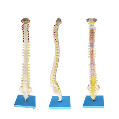
Secondly, the soft silicone anatomical model has the advantages of three-dimensional and high simulation, which can reproduce the three-dimensional structure and entity information of tissues and organs. The arteries (red), veins (blue) and nerves (yellow) in the medical soft silicone anatomical model products are formed by integrating color separation filling and perfusion, which is not a conventional staining method and does not fade. Muscle organs and other parts according to the color of perfusion molding.
Thirdly, The soft silicone anatomy model changes the traditional anatomy teaching mode, overcomes the shortcomings of monotonous, rigid and too abstract teaching, and stimulates students' learning initiative and enthusiasm.
According to the needs of teaching scenes and links, soft silicone anatomical model is used as teaching AIDS, combined with imaging data and traditional anatomical maps, so that students can understand and master the professional knowledge points of anatomy more easily, which not only improves the teaching quality and efficiency, but also makes teaching and learning easy and pleasant.
Do you know how to make soft silicone anatomical model for medical education? Meiwo will take you through the production process of soft silicone anatomical models.
First, sculpture & shaping
Combined with the current domestic latest medical teaching materials and the latest atlas of international authorities, according to the needs of customers, or doctors and students to create more subtle and unique realistic products, molding artists use food grade fine sculpting oil clay sculpting. Environmental clay sculpting model details, lifelike.
Second, mold manufacturing
Deployment of environmental protection silicone, in the fine carved modeling, repeated silicone production, or covered with gauze to strengthen the mold, and finally plaster fixed mold molding.
Third, painting & rapid solidification
Adjust food-grade silicone gel according to the proportion, fill color painting in the corresponding mold parts according to the different colors of each organ on the produced mold, divide the layers, fill color layer by layer, maintain the corresponding thickness, and proceed to the next step after curing.
Fourth, fill with foaming agent
Pour an appropriate amount of foaming agent into the prototype mold of the product, and the product will be lighter and have texture, easy to teach and learn.
Fifth, compound die & edge sealing
After foaming, silicone rubber is poured into the product mold, and the product mold is fitted up and down, fixed with plaster, trimmed and beautified. The product production is completed, and the finished product is environmentally friendly, practical and beautiful.
The material of soft silicone model products is mainly made of silicone rubber and its auxiliary chemical products. The ROHS certification is the safest and environmentally friendly chemical material in the rubber and plastic industry at present. Moreover, the soft silicone model products have been certified by FDA and meet the environmental protection standards, which can be used in the field of food and cosmetics. So soft silicone model products are non-toxic and tasteless. And the production of soft silicone model products using silicone different degrees of hardness to reflect different organs of the human body, so that the soft silicone model can more directly reflect the internal structure of human organs; The image is more realistic.
The main material of PVC model products is made of PVC powder, oil and color paste mixed and blended. The raw materials are heated and cooled by micro-injection glue dropping process. PVC soft glue has the difference between ordinary material and environmental protection material, environmental protection material is also called ATBC-PVC, can pass some lower standards of environmental protection certification, but there is also some toxicity, can not be used in the field of food and cosmetics. PVC model products are generally hard in texture, which can only simply reflect the shape of human tissues, but can not directly and objectively express the degree of softness and hardness of different human tissues.
Soft silicone model products feel soft, good toughness and elasticity, smooth appearance, color, brightness. Because the silicone material has many other similar materials difficult to replace the characteristics, its high adsorption performance, good thermal stability, chemical stability, and high mechanical strength, so the production of the model is durable, the surface of the product can not see the pores, with smooth and realistic feel and accurate shape, convenient teaching display and simulation anatomy training, It is very scientific and practical.


Human Anatomy Models
No discipline of science relies more on the use of anatomical models than human anatomy and physiology. Preserved human tissues are not readily available for educational purposes, and when they are, the condition of the specimens is usually not satisfactory to show detailed morphology. Many structures, such as the components of the middle and inner ear, are much too small for convenient study from natural specimens.
Human torso models
A complete human figure or torso model with head is actually a collection of models of individual organs, many of which can be separated and even opened for internal inspection. A human torso model of high quality is a valuable acquisition for any anatomy classroom. Generally, those with the largest number of removable components have the greatest teaching value.
Skeleton models
A staple in many science classrooms, skeletons allow students to examine bones without other organs obstructing their view. Disarticulated skeletons are perfect for learning about the shapes and structure of bone; they are also great for forensic studies. Muscular skeletons have origin and insertion points indicated on the bones to help students understand how the skeletal and muscular systems work together to gain an appreciation for movement. Flexible and ligamentous skeletons offer students an opportunity to visualize the movement of the skeletal system and the articulation of joints.
Organ and organ system models
For a detailed look at specific organs and structures, consider organ and organ system models. These provide a more focused view of the major anatomical structures within systems like the respiratory and urinary systems. Organ models are often made of many pieces, making it easy to explore the internal structures as well.
Disease and pathology models
Providing examples of disease and pathology for students is paramount to understanding the structure and function of human tissue. Tissue pathology models give students an opportunity to observe enlarged examples of diseased tissue, with aids in their ability to clearly identify similar features under a microscope. Organ pathology and disease models, including this hypertension model, show how disease affects the entire system or body.
Care of models
Models represent a formidable investment, and with reasonable care, they will last for many years. Encourage your students to handle the models, but to do so with care and respect, particularly when removing or replacing parts. Breakage occurs most frequently when parts of models are forced together or twisted apart. It is especially important that students do not touch the models with pens or pencil tips, because stains are difficult to remove without damaging the paint.
Soft Silicone Anatomy Models
Shoulder muscles
The shoulder muscle is located around the shoulder joint, can move the shoulder joint, and enhance the stability of the shoulder joint, including the deltoid, shoulder spleen inferior, supraspinatus, infraspinatus, teres major, teres minor.
Brachial muscle
The arm muscles can be divided into anterior (flexor) and posterior (extensor) groups.
The biceps brachialis is located in front of the arm, the muscle abdomen is fusiform, with long and short ends. Function of bending the elbow joint, so that the forearm supination, long head can also help to bend the shoulder joint.
The posterior triceps are located behind the upper arm and have 3 starting heads, namely the long head, the medial head and the lateral head. Function to extend the elbow joint, long head can make the arm back extension and adduction.
Forearm muscles
The forearm muscles are located around the burning and ulna bones. There are 19 muscles in total, divided into anterior and posterior groups.
The anterior group is located in the front of the forearm, mainly for wrist flexion, flexion and forearm pronation muscles, so it is called flexor muscle group, a total of 9 muscles, divided into shallow and deep two layers.
The posterior group is located at the back of the forearm, and its main function is to extend the wrist, extend the fingers and pronate the forearm, so it is called the extensor muscle group, a total of 11 muscles, divided into shallow and deep layers arranged.
Hand muscles
All are located in the palm of the hand, divided into the lateral group, the middle group and the medial group, the main role for the movement of the fingers.
dissection muscle of human upper limb anatomy model shows that: the motor and venous nerves of the superficial and deep layers of the upper limbs can be separated from the deltoid muscle, biceps brachii, triceps brachii, palmtop muscle, flexor carpi radialis, extensor carpi brachii, and arpal arpal musculi to show the shape, position, proximity, starting and ending points of the upper limbs muscles. Subclavian artery branch, brachial artery branch, ulnar artery branch, radial artery branch good deep arch, superficial arch, upper limb ulnar nerve, radial nerve distribution, the main venous return of the upper limb, etc.
Meiwo science software products cover classroom teaching, experimental teaching, and life science museum display system. Specifically, there are multiple sets of medical software such as comprehensive teaching platform for human anatomy, digital human anatomy system, 3D specimen database query system, human body nine interactive display system, VR human anatomy, and assembly of human organs, etc.
Meiwo adopts the construction concept of "intelligent life science museum", which integrates the intelligent management of the exhibition hall, the professional display of specimens and the scientific display of medical knowledge.The exhibition hall will provide a high-tech, intelligent and experiential interactive learning platform with the combination of human experimental teaching, popular science education, clinical application, humanistic care, cultural inheritance, medicine and art, etc.

Q: What is soft silicone used for?
Q: What are the uses of anatomical models?
Q: What are 3D printed anatomical models?
Q: What material is used to make anatomical models?
Q: Is soft silicone body safe?
Q: What does silicone do to the body?
Q: What is the difference between soft silicone and liquid silicone?
Q: What are soft silicone anatomy models?
Q: What material is used for prosthetic bones?
Q: What is the best prosthesis for hip replacement?
Q: Is silicone soft like rubber?
Q: How are soft silicone anatomy models made?
Q: Are these models suitable for veterinary training?
Q: Are there ethical concerns with using soft silicone models?
Q: Do they provide a realistic simulation for all medical procedures?
Q: Can they be customized for specific educational needs?
Q: Do they come in various sizes and anatomical details?
Q: Do they offer a cost-effective solution for educational institutions?
Q: How do they compare to digital or virtual models?
Q: Do they require special care or maintenance?