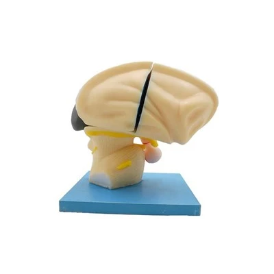
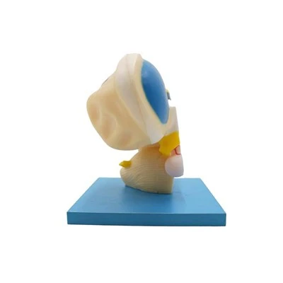
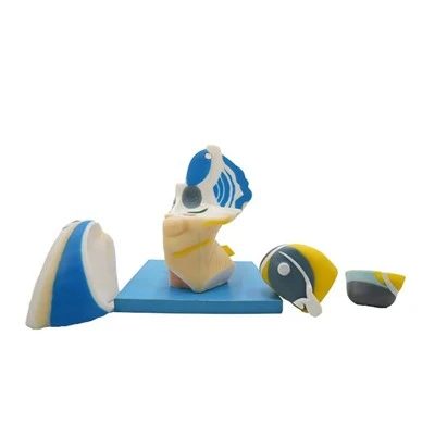
Brain model parts, enlarged diencephalon, telencephalon, thalamus for medical education was made of environmental silicone, shows the structure of the diencephalon and part of the telencephalon.
The diencephalon, telencephalon, and thalamus are all important brain model parts. In anatomy teaching, models of these organs are crucial for understanding their structure and positional relationships.
The diencephalon is located between the midbrain and telencephalon, mostly covered by the cerebral hemispheres. It mainly comprises five parts: the dorsal thalamus, hypothalamus, epithalamus, subthalamus, and posterior thalamus. The internal cavity of the diencephalon is the third ventricle. In anatomical models, the diencephalon is usually designed to show its morphology covered by the cerebral hemispheres and its complex internal nuclei.
The telencephalon, commonly referred to as the brain, is mainly composed of the left and right cerebral hemispheres. The surface of the cerebral hemispheres is covered with grooves and fissures of varying depths; the ridges between the shallow grooves are called gyri. The thalamus, part of the diencephalon, is located dorsally to the diencephalon and consists of a pair of oval-shaped gray matter masses, accounting for approximately 4/5 of the diencephalon's volume.
The thalamus has a complex internal structure, containing three major nuclei: the anterior nuclei, medial nuclei, and lateral nuclei. In anatomical models, thalamic models focus on demonstrating the distribution of its internal nuclei and their connections to other brain regions.
Brain Model Parts, Enlarged Diencephalon, Telencephalon, Thalamus Details:
Product Name
Brain Model Parts, Enlarged Diencephalon, Telencephalon, Thalamus
Description
The structure of the diencephalon and part of the telencephalon, the relative relationship of the hypothalamus kernel mass were demonstrated in detail, and the dorsal thalamus was designed to be detachable.
Size
Enlarged, 25x18x25cm
Material
Environmental protection soft silicone and paint
Application
Medical teaching exhibition and display science research science education
MOQ
1
Meiwo Service:
High quality products and professional services have always been the foundation of meiwo's survival.
Pre-sale service
Product and publicity picture design
Exhibition & museum layout
Technical support
All the silicone anatomy models and animal plastinated models can also be customized according to the requirements.
After-sale service
1. The warranty time is 2 years from the day of receiving goods day.
2. After 2 years warranty, only charge cost of maintenance and replacement.

 Language